Cross Section Of A Bone : Medullary cavity - Wikipedia
Cross Section Of A Bone : Medullary cavity - Wikipedia. This is known as the periosteum. They are obtained by taking imaginary slices perpendicular to the main axis of organs, vessels, nerves, bones, soft tissue, or even the entire human body. Browse 53 bone marrow cross section stock photos and images available, or search for bone cross section or bone cells to find more great stock photos and pictures. Two types of bone tissues in cross section of a long bone : The cortical bone equivalent area of the cross‐section of the region of interest (femoral neck or shaft), with all soft tissue voids (trabecular and cellular spaces) eliminated (cm 2).
ads/bitcoin1.txt
Start studying cross section of long bone. Human bone, cross section diagram of femur showing osteon, veins, marrow. This slide contained a cross section of a very small bone, and you are looking at the entire thickness of the shaft of the bone. Internal structure of a human long bone, with a magnified cross section of the interior. There are three general classes of bone.
Is it soft and a flabby under the feet of an elephant? - Quora from qph.fs.quoracdn.net Find the perfect cross section bone stock photo. Huge collection, amazing choice, 100+ million high quality, affordable rf and rm images. While it is not as hard as compact bone, spongy bone plays an important role of protecting the marrow where blood cells are produced. Compact bone is the outer layer and the spongy bone forms the inner layer. Slides have to be made this way because the matrix of bone is too hard to be cut with a knife as the other tissues are. If you look at the cross section of a long bone under a microscope, the rings of bone immediately internal to the periosteum of the bone are called external circumferential lamellae. Two types of bone tissues in cross section of a long bone : Bone markings the surface features of bones vary considerably, depending on the function and location in the body.
100x first focus in the compact decalcified bone (cb) on the left part of the image, you can see small dots, which are.
ads/bitcoin2.txt
Start studying cross section of long bone. This is known as the periosteum. Select from premium cross section of bone of the highest quality. Learn vocabulary, terms, and more with flashcards, games, and other study tools. The diaphysis is the tubular shaft that runs between the proximal and. Huge collection, amazing choice, 100+ million high quality, affordable rf and rm images. Explaned distal and proximal epiphysis. There are three general classes of bone. As the names suggest compact bone looks compact and the spongy bone looks like sponges. And why does the marrow stop where it does, and so sharply? Beautiful tooth cross section illustration, deep blue background and sparkling lights around. There are trabeculae in spongy bone which gives its sponge like appearance. This slide contained a cross section of a very small bone, and you are looking at the entire thickness of the shaft of the bone.
The cortical bone equivalent area of the cross‐section of the region of interest (femoral neck or shaft), with all soft tissue voids (trabecular and cellular spaces) eliminated (cm 2). The upper (biting) surfaces of the tooth are at top, with the lower sections (bottom) embedded in the gums and jaw bone (not shown). The diaphysis and the epiphysis. Beautiful tooth cross section illustration, deep blue background and sparkling lights around. The structure of a long bone allows for the best visualization of all of the parts of a bone.
An Inside Look | Smithsonian National Museum of Natural ... from naturalhistory.si.edu Related posts of cross section of a long bone bone structure right foot. Find the perfect cross section bone stock photo. Related posts of cross section of human bone diagram muscles and bones of the human body. After a fracture, woven bone forms initially and is gradually replaced by lamellar bone during a process known as bony substitution. Cross‐sectional area is derived from the integral of the bone mass profile across the narrow region. They are obtained by taking imaginary slices perpendicular to the main axis of organs, vessels, nerves, bones, soft tissue, or even the entire human body. Now that you know what bones do, let's take a look at what they're made of and their anatomy. There are three general classes of bone.
Compact bone, spongy bone, and bone marrow.
ads/bitcoin2.txt
A long bone has two parts: At the end of the bone is the epiphysis, which in young people is separated from the. 100x first focus in the compact decalcified bone (cb) on the left part of the image, you can see small dots, which are. Cross‐sectional area is derived from the integral of the bone mass profile across the narrow region. Two types of bone tissues in cross section of a long bone : Bone markings the surface features of bones vary considerably, depending on the function and location in the body. The cortical bone equivalent area of the cross‐section of the region of interest (femoral neck or shaft), with all soft tissue voids (trabecular and cellular spaces) eliminated (cm 2). Vector illustration scheme of bone cross section. Compact bone is the outer layer and the spongy bone forms the inner layer. Start studying cross section of long bone. And why does the marrow stop where it does, and so sharply? The upper (biting) surfaces of the tooth are at top, with the lower sections (bottom) embedded in the gums and jaw bone (not shown). The compact bone is made up of osteon.
After a fracture, woven bone forms initially and is gradually replaced by lamellar bone during a process known as bony substitution. The central tubular region of the bone, called the diaphysis, flares outward near the end to form the metaphysis, which contains a largely cancellous, or spongy, interior. 100x first focus in the compact decalcified bone (cb) on the left part of the image, you can see small dots, which are. Compact bone is the outer layer and the spongy bone forms the inner layer. The large dark spots are passages for blood vessels and nerves.
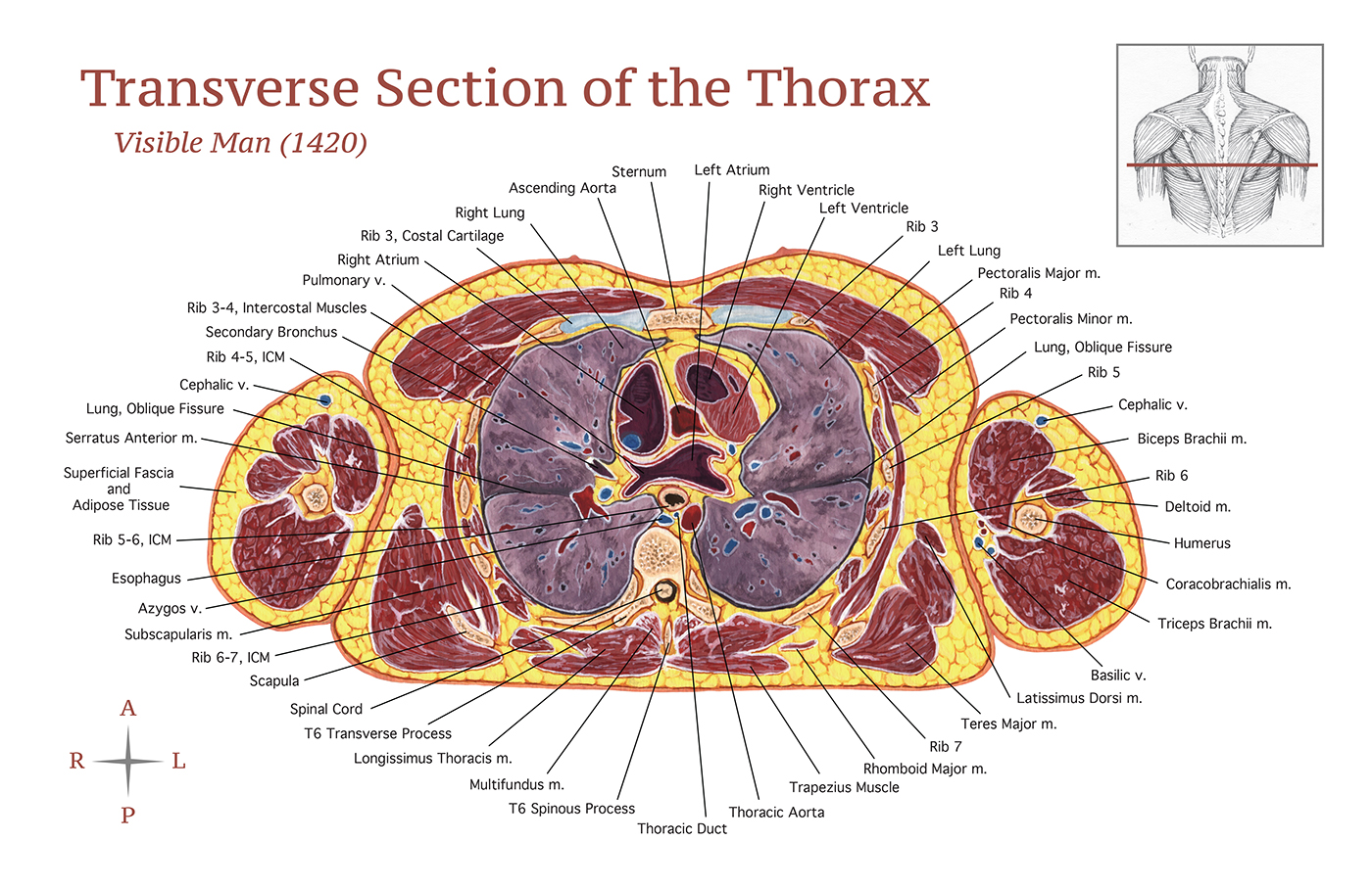
Transverse Section of the Thorax on Behance from mir-s3-cdn-cf.behance.net Cross‐sectional area is derived from the integral of the bone mass profile across the narrow region. Diagram with articular cartilage, marrow, spongy bone, medullary cavity, endosteum, diaphysis, and periosteum. Browse 4,246 bone cross section stock photos and images available, or search for human bone cross section to find more great stock photos and pictures. As the names suggest compact bone looks compact and the spongy bone looks like sponges. The large dark spots are passages for blood vessels and nerves. Learn vocabulary, terms, and more with flashcards, games, and other study tools. Vector illustration scheme of bone cross section. A long bone has two parts:
Two types of bone tissues in cross section of a long bone :
ads/bitcoin2.txt
This is known as the periosteum. And why does the marrow stop where it does, and so sharply? Select from premium cross section of bone of the highest quality. I don't find it enhances the image. The large dark spots are passages for blood vessels and nerves. There are three general classes of bone. 100x first focus in the compact decalcified bone (cb) on the left part of the image, you can see small dots, which are. Compact bone, spongy bone, and bone marrow. The surface features of bones vary considerably, depending on the function and location in the body. Marrow in the shaft of long bones is typically yellow, with red marrow in the head through the cancellous bone. Diagram with articular cartilage, marrow, spongy bone, medullary cavity, endosteum, diaphysis, and periosteum. Why is the marrow red? Vector illustration scheme of bone cross section.
ads/bitcoin3.txt
ads/bitcoin4.txt
ads/bitcoin5.txt
0 Response to "Cross Section Of A Bone : Medullary cavity - Wikipedia"


0 Response to "Cross Section Of A Bone : Medullary cavity - Wikipedia"
Post a Comment